missing translation for 'onlineSavingsMsg'
Learn More
Learn More


Invitrogen™ CD279 (PD-1) Monoclonal Antibody (J121), eBioscience™
Mouse Monoclonal Antibody
504.00€
Spécification
| Antigène | CD279 (PD-1) |
|---|---|
| Clone | J121 |
| Concentration | 0.5 mg/mL |
| Contenu et stockage | 4°C |
| Applications | Immunohistochemistry (Paraffin), Western Blot, Immunocytochemistry |
Description
Description: This J121 monoclonal antibody reacts with human CD279 (programmed death-1, PD-1), a 55 kDa member of the CD28 immunoglobulin superfamily. CD279 contains the immunoreceptor tyrosine-based inhibitory motif (ITIM) and plays a key role in peripheral tolerance and autoimmune disease. CD279 is expressed predominantly on activated T and B lymphocytes. Two novel members of the B7 family have been identified as the CD279 ligands, CD274 (PD-L1, B7-H1) and CD273 (PD-L2, B7-DC). Evidence reported to date suggests overlapping functions for these two ligands and their constitutive expression on some normal tissues and upregulation on activated antigen-presenting cells. More recently, therapies targeting the blockade of the CD279/CD274 pathway have become the focus for treatment of melanoma, renal cell cancer, Hodgkins' lymphoma, and non-small cell lung carcinoma (NSCLC). The J121 monoclonal antibody is not recommended for flow cytometry of human cells. For detection of human CD279 using flow cytometry please refer to clone eBioJ105 (J105). Applications Reported: This J121 antibody has been reported for use in western blotting, and immunohistochemical staining of formalin-fixed paraffin embedded tissue sections. Applications Tested: This J121 antibody has been tested by immunohistochemistry of formalin-fixed paraffin embedded human tissue using high or low pH antigen retrieval and can be used at less than or equal to 5 μg/mL. It is recommended that the antibody be care...
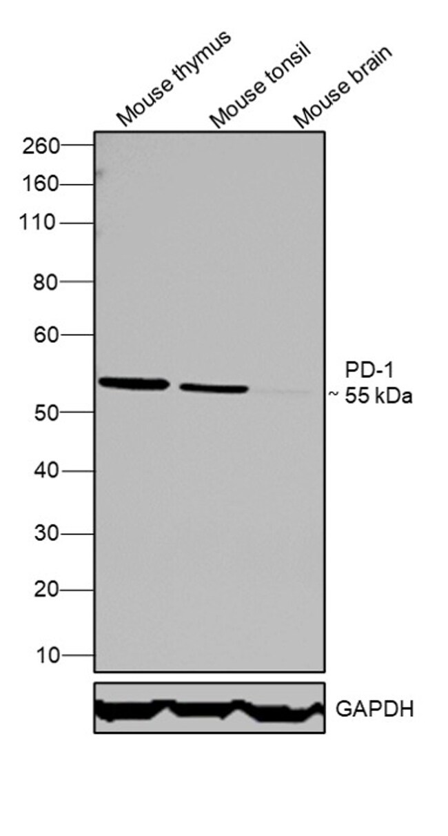
Cell-mediated immune responses are initiated by T lymphocytes that are themselves stimulated by cognate peptides bound to MHC molecules on antig en-presenting cells (APC). T-cell activation is generally self-limited as activated T cells express receptors such as PD-1 (also known as PDCD-1) that mediate inhibitory signals from the APC. PD-1 can bind two different but related ligands, PDL-1 and PDL-2. Upon binding to either of these ligands, signals generated by PD-1 inhibit the activation of the immune response in the absence of 'danger signals' such as LPS or other molecules associated with bacteria or other pathogens. Evidence for this is seen in PD1-null mice who exhibit hyperactivated immune systems and autoimmune diseases. Despite its predicted molecular weight, PD-1 often migrates at higher molecular weight in SDS-PAGE.Spécification
| CD279 (PD-1) | |
| 0.5 mg/mL | |
| Immunohistochemistry (Paraffin), Western Blot, Immunocytochemistry | |
| Unconjugated | |
| Mouse | |
| RUO | |
| PBS with 0.09% sodium azide; pH 7.2 | |
| Q02242, Q15116 | |
| 18566, 5133 | |
| Primary | |
| Affinity chromatography |
| J121 | |
| 4°C | |
| Monoclonal | |
| Liquid | |
| IgG1 κ | |
| Human, Mouse | |
| Pdcd1 | |
| CD279; EGK_05005; hPD1; hPD-1; hPD-l; hSLE1; Ly101; mPD-1; PD1; PD-1; Pdc1; Pdcd1; programmed cell death 1; programmed cell death 1 protein; programmed cell death protein 1; programmed cell death protein 1-like; programmed death 1; Protein PD1; protein PD-1; sCD279; SLEB2; soluble CD279; systemic lupus erythematosus susceptibility 2 | |
| Pdcd1 | |
| Antibody |
Vous avez repéré une opportunité d'amélioration ?Partager une correction de contenu
Correction du contenu d'un produit
Veuillez fournir vos retours sur le contenu du produit en remplissant le formulaire ci-dessous.
Nom du produit