missing translation for 'onlineSavingsMsg'
Learn More
Learn More


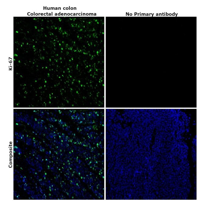
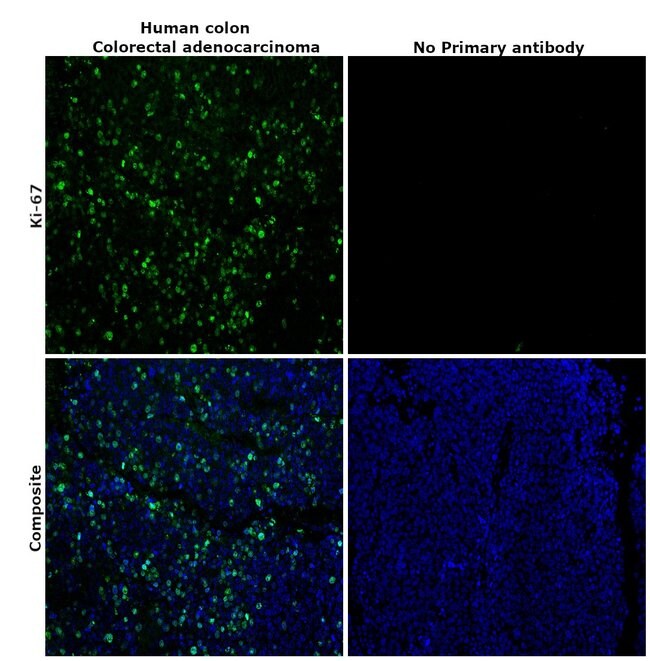
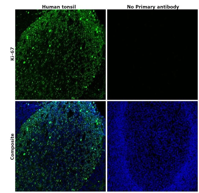
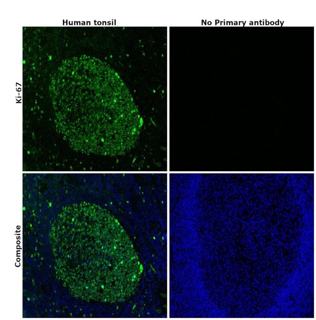
Invitrogen™ Ki-67 Monoclonal Antibody (SolA15), Alexa Fluor™ 488, eBioscience™, Invitrogen™
Rat Monoclonal Antibody
173.00€ - 401.00€
Spécification
| Antigène | Ki-67 |
|---|---|
| Clone | SolA15 |
| Concentration | 0.5 mg/mL |
| Applications | Flow Cytometry, Immunohistochemistry (Paraffin), Immunocytochemistry |
| Classification | Monoclonal |
| Code produit | Marque | Quantité | Prix | Quantité et disponibilité | |||||
|---|---|---|---|---|---|---|---|---|---|
| Code produit | Marque | Quantité | Prix | Quantité et disponibilité | |||||
|
16310994
|
Invitrogen™
53-5698-80 |
25 μg |
173.00€
25µg |
Veuillez vous connecter pour pouvoir commander cet article. Besoin d'un compte web? Créer le vôtre dès maintenant! | |||||
|
16320994
|
Invitrogen™
53-5698-82 |
100 μg |
401.00€
100µg |
Veuillez vous connecter pour pouvoir commander cet article. Besoin d'un compte web? Créer le vôtre dès maintenant! | |||||
Description
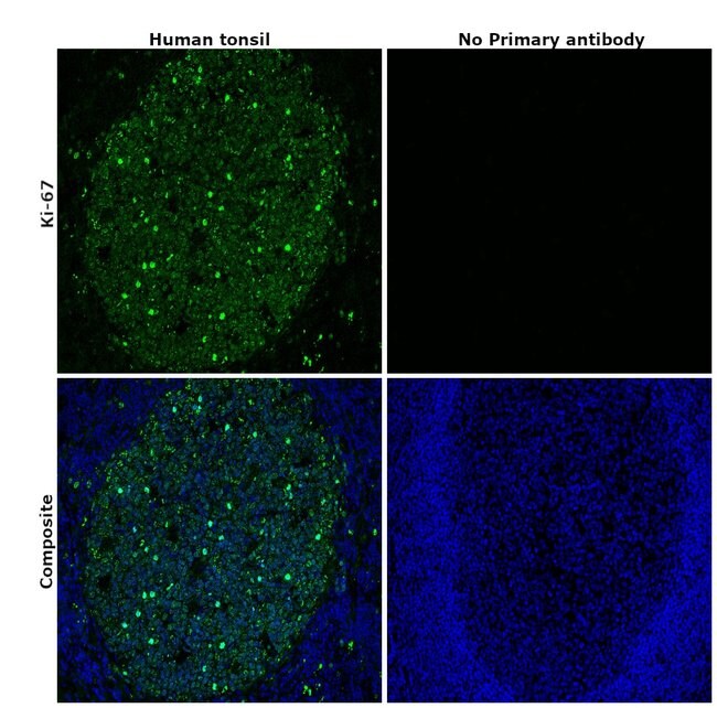
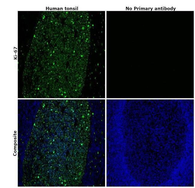
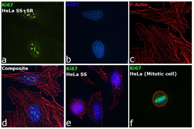
Description: The monoclonal antibody SolA15 recognizes mouse and rat Ki-67, a 300 kDa nuclear protein. Ki-67 is present during all active phases of the cell cycle (G1, S, G2, and mitosis), but is absent from resting cells (G0). Ki-67 is detected within the nucleus during interphase but redistributes to the chromosomes during mitosis. Ki-67 is used as a marker for determining the growth fraction of a given population of cells. In studies of tumor cells, the Ki-67 labeling index refers to the number of Ki-67 positive cells within the population and this is used to predict outcome of particular cancer types. Ki-67 has been shown to interact with the DNA-bound protein chromobox protein homolog 3 (CBX3) (heterochromatin). The SolA15 antibody also recognizes human, non-human primate and canine Ki-67. Applications Reported: This SolA15 antibody has been reported for use in intracellular staining followed by flow cytometric analysis. Applications Tested: This SolA15 antibody has been tested by intracellular staining and flow cytometric analysis of mouse splenocytes using the Foxp3/Transcription Factor Staining Buffer Set (cat. 00-5523) and protocol. Refer to Best Protocols for Staining Protocol (refer to Protocol B: One-step protocol for intracellular (nuclear) proteins). This can be used at less than or equal to 0.25 μg per test. A test is defined as the amount (μg) of antibody that will stain a cell sample in a final volume of 100 μL.
Ki-67 is a nuclear protein that is expressed during various stages in the cell cycle, particularly during late G1, S, G2, and M phases. The protein has a forkhead associated domain (FHA) through which it associates with euchromatin at the perichromosomal layer, the centromeric heterochromatin, and the nucleolus. Ki-67 is shown to have a cell cycle dependent topographical distribution with perinucleolar expression at G1, expression in the nuclear matrix at G2, and expression on the chromosomes during M phase. Ki-67 is commonly used as a proliferation marker because it is not detected in G0 cells, but increases steadily from G1 through mitosis. Ki-67 antibodies are useful in establishing the cell growing fraction in neoplasms. In neoplastic tissues, the prognostic value is comparable to the tritiated thymidine-labelling index. The correlation between low Ki-67 index and histologically low-grade tumors is strong. Ki-67 is routinely used as a neuronal marker of cell cycling and proliferation.Spécification
| Ki-67 | |
| 0.5 mg/mL | |
| Monoclonal | |
| Liquid | |
| RUO | |
| PBS with 0.09% sodium azide; pH 7.2 | |
| antigen identified by monoclonal antibody Ki 67; antigen identified by monoclonal antibody Ki-67; Antigen identified by monoclonal antibody Ki-67 homolog; Antigen KI-67; Antigen KI-67 homolog; antigen KI-67; proliferation marker protein Ki-67; antigen KI-67-like; cb31; D630048A14Rik; I79_022666; Ki67; Ki-67; KIA; LOW QUALITY PROTEIN: proliferation marker protein Ki-67; marker of proliferation Ki-67; MIB-; MIB-1; Mki67; PPP1R105; Proliferation marker protein Ki-67; proliferation-related Ki-67 antigen; protein phosphatase 1, regulatory subunit 105; RP11-380J17.2; sb:cb31; si:ch211-250b22.7; unnamed protein product; wu:fa11g09; wu:fb57a07; wu:fi14e05 | |
| Mki67 | |
| Primary | |
| 4°C, store in dark, DO NOT FREEZE! | |
| Mki67 |
| SolA15 | |
| Flow Cytometry, Immunohistochemistry (Paraffin), Immunocytochemistry | |
| Alexa Fluor 488 | |
| Rat | |
| Canine, Cynomolgus Monkey, Human, Mouse, Monkey, Rat | |
| E9PVX6, P46013 | |
| 100686578, 102135895, 17345, 291234, 4288 | |
| IgG2a κ | |
| Affinity chromatography | |
| Antibody |
Vous avez repéré une opportunité d'amélioration ?Partager une correction de contenu
Correction du contenu d'un produit
Veuillez fournir vos retours sur le contenu du produit en remplissant le formulaire ci-dessous.
Nom du produit